Leg Muscles Diagram / Muscles Of The Leg And Foot Classic Human Anatomy In Motion The Artist S Guide To The Dynamics Of Figure Drawing. The biceps femoris is a muscle of the posterior thigh composed of a long head and a short head. The calf muscle, on the back of the lower leg, is actually made up of two muscles: On the medial edge of the posterior thigh is the gracilis muscle. Related posts of lower leg muscles diagram muscle anatomy neck. For images of the muscle, click on each link under location.
The muscles in the hip are responsible for the movement of the hip and, by proxy, the leg. The biceps femoris is a muscle of the posterior thigh composed of a long head and a short head. These muscles are found on the front and back sides of the lower leg. Legs are used for standing, and all forms of. Flexes elbow and moves forearm.
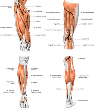
13 599 Best Leg Muscles Anatomy Images Stock Photos Vectors Adobe Stock from t4.ftcdn.net The largest muscle masses in the leg are present in the thigh and the calf. This is why you have to indicate which biceps you are taking about when discussing one or other of these muscles. In the leg muscles diagram above, there are many muscles that make up your legs and support it to move. Observe the leg muscle diagram posted above and notice that there are many parts in the muscles. There are many muscles located in the lower leg, but there are three that are particularly well known—the gastrocnemius and the soleus, which are the most powerful muscles in the lower leg, and the anterior tibialis. The calf muscle, on the back of the lower leg, is actually made up of two muscles: Extends spine and trunk back. Muscle of the human leg diagram in this image, you will find muscle of the human leg diagram, hip and femur middle layer, hip and femur deep layer, overview of the most important muscles of the leg, femur middle layer, femur deep layer, rectus femoris m.
It is also visible on the medial edge of the thigh from the anterior.
Supporting, balancing, and propelling the body is the work of the muscular system of the legs and feet. Take a look at the leg muscles diagram below, where you see each muscle clearly labeled. These muscles are found on the front and back sides of the lower leg. There are many muscles located in the lower leg, but there are three that are particularly well known—the gastrocnemius and the soleus, which are the most powerful muscles in the lower leg, and the anterior tibialis. Human anatomy diagrams show internal organs, cells, systems, conditions, symptoms and sickness information and/or tips for healthy living. The long head arises from a common tendon with semitendinosus from the superior medial quadrant of the posterior portion of the ischial tuberosity. The muscles work together to enable movement and keep the hip in alignment. The muscles in the front allow for. Notice the upper leg has a biceps muscle just like the upper arm does. The quad muscles— which form the meaty mass on the front of your thighs — are among your strongest muscle groups, and play a critical role in athletic activities. This is the group of muscles that you often see body builders flexing, which protrude just above the knee and take up most of the upper leg. The 3 muscles are called triceps coxae. Spend some time revising this diagram by connecting the name and location of each structure with what you've just learned in the video.
This is why you have to indicate which biceps you are taking about when discussing one or other of these muscles. The hamstring muscles, also known as the rear thighs, make up the backside of the upper leg anatomy. Spend some time revising this diagram by connecting the name and location of each structure with what you've just learned in the video. It is also visible on the medial edge of the thigh from the anterior. The muscles that make up the quadriceps are the strongest and leanest of all muscles in the body.
Impact Of Loss Of Wing Muscles On Geometry Of Leg Muscles And Petiole Download Scientific Diagram from www.researchgate.net The quad muscles— which form the meaty mass on the front of your thighs — are among your strongest muscle groups, and play a critical role in athletic activities. The muscles in the front allow for. See more ideas about muscle anatomy, leg muscles anatomy, leg muscles. Extends spine and trunk back. This is the group of muscles that you often see body builders flexing, which protrude just above the knee and take up most of the upper leg. The femoral, saphenous, obturator, and lateral femoral cutaneous nerves all extend from the lumbar plexus into the muscles and skin of the thigh and leg. The 3 muscles are called triceps coxae. Observe the leg muscle diagram posted above and notice that there are many parts in the muscles.
One of the most important tendons in terms of mobility of the leg is the achilles tendon.
Like the quadriceps, the hamstring muscle group also contains four separate muscles: The hamstring muscles, also known as the rear thighs, make up the backside of the upper leg anatomy. Extends spine and trunk back. The muscles in the hip are responsible for the movement of the hip and, by proxy, the leg. Your quadricep muscles, also known as quads, consist of four muscles that compose the front of your leg; This diagram depicts anatomy of leg muscles. This important tendon in the back of the calf and ankle stores the elastic energy needed for running, jumping, and other physical activity. Take a look at the leg muscles diagram below, where you see each muscle clearly labeled. A muscle located on the back portion of the lower leg, being one of the two major muscles that make up the calf:the flexing of this muscle during walking and bending of the knee creates traction on the femur, pulling it toward the tibia in the lower leg and causing the knee to bend. It is also visible on the medial edge of the thigh from the anterior. The human leg, in the general word sense, is the entire lower limb of the human body, including the foot, thigh and even the hip or gluteal region. See more ideas about muscle anatomy, human anatomy and physiology, body anatomy. The 3 muscles are called triceps coxae.
Related posts of lower leg muscles diagram muscle anatomy neck. Muscles of the leg and foot. Observe the leg muscle diagram posted above and notice that there are many parts in the muscles. The 3 muscles are called triceps coxae. For women, shaping the thigh muscles is an essential goal of physical fitness.
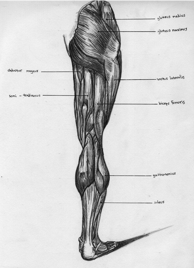
Leg Back Muscle Chart By Badfish81 On Deviantart from images-wixmp-ed30a86b8c4ca887773594c2.wixmp.com Muscles of the leg and foot. The gastrocnemius muscle has two large bellies, called the medial head and the lateral head, and inserts into the calcaneus bone of the foot via its calcaneal tendon (also known as the achilles tendon.) The aim of this exercise is to improve your confidence in identifying different structures. Kegel muscle anatomy 12 photos of the kegel muscle anatomy kegel muscle anatomy, human muscles, kegel muscle anatomy. The calf muscle, on the back of the lower leg, is actually made up of two muscles: Anterior compartment thigh muscles this is the largest of the three compartments of the thigh. It is also visible on the medial edge of the thigh from the anterior. The biceps femoris is a muscle of the posterior thigh composed of a long head and a short head.
The calf muscle, on the back of the lower leg, is actually made up of two muscles:
Together, these muscles straighten your knee, stabilize your knee joint, assist in flexing your hip (drawing your knee towards your chest), and help absorb force when you land after jumping or leaping. Related posts of muscles and tendons of the leg kegel muscle anatomy. Biceps femoris (long head) biceps femoris (short head) semitendinosus. The following diagram illustrates the actions of the terms adduction, abduction, flexion and extension at the different joints. This is the group of muscles that you often see body builders flexing, which protrude just above the knee and take up most of the upper leg. Notice the upper leg has a biceps muscle just like the upper arm does. Raises and rotates arm in all directions. The biceps femoris is a muscle of the posterior thigh composed of a long head and a short head. Brings hip away from body. Human anatomy diagrams show internal organs, cells, systems, conditions, symptoms and sickness information and/or tips for healthy living. Kegel muscle anatomy 12 photos of the kegel muscle anatomy kegel muscle anatomy, human muscles, kegel muscle anatomy. It is also visible on the medial edge of the thigh from the anterior. The long head arises from a common tendon with semitendinosus from the superior medial quadrant of the posterior portion of the ischial tuberosity.